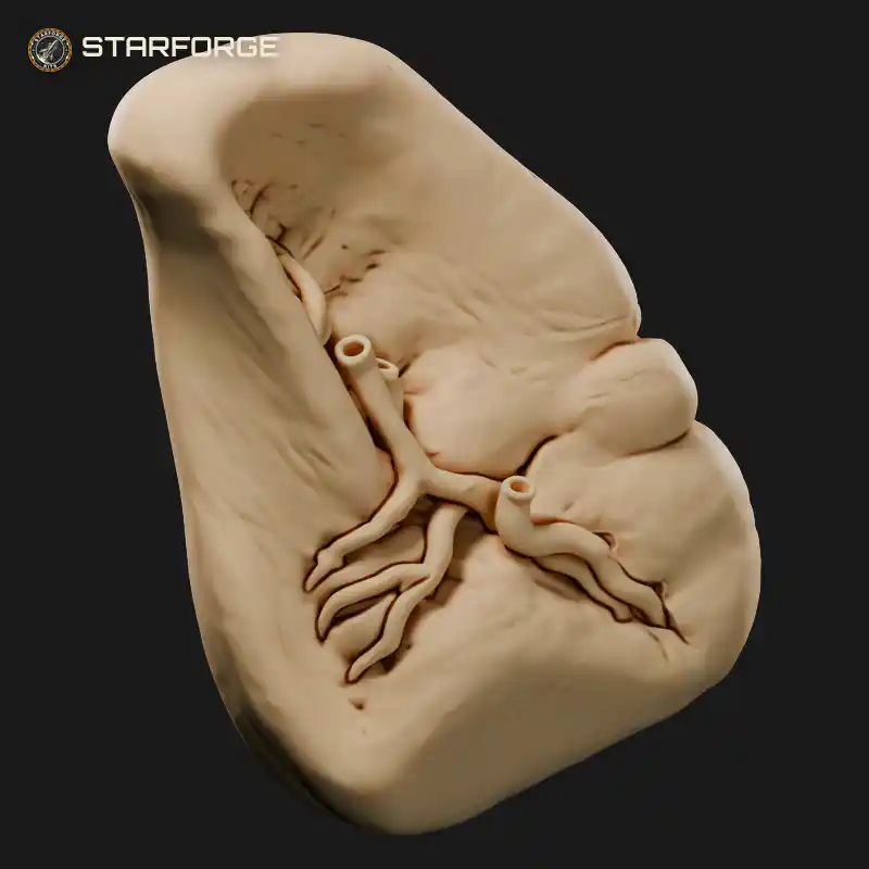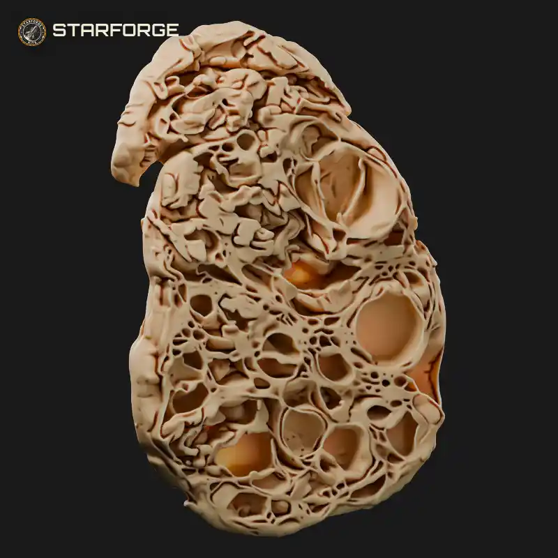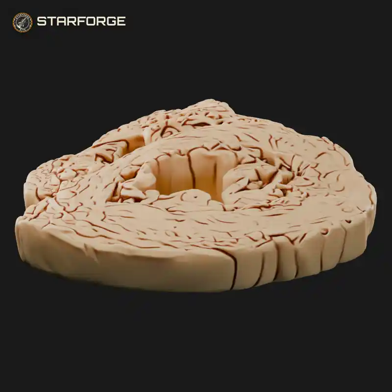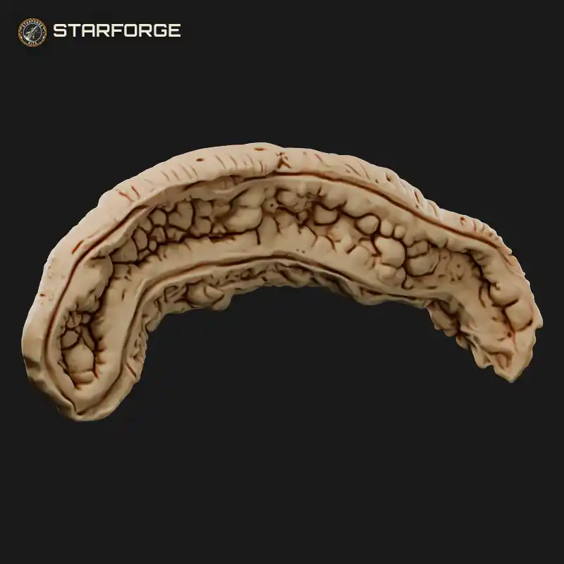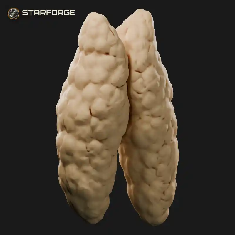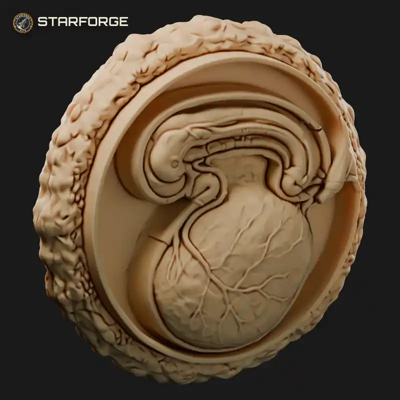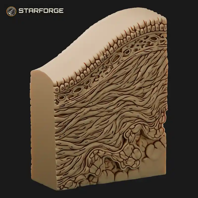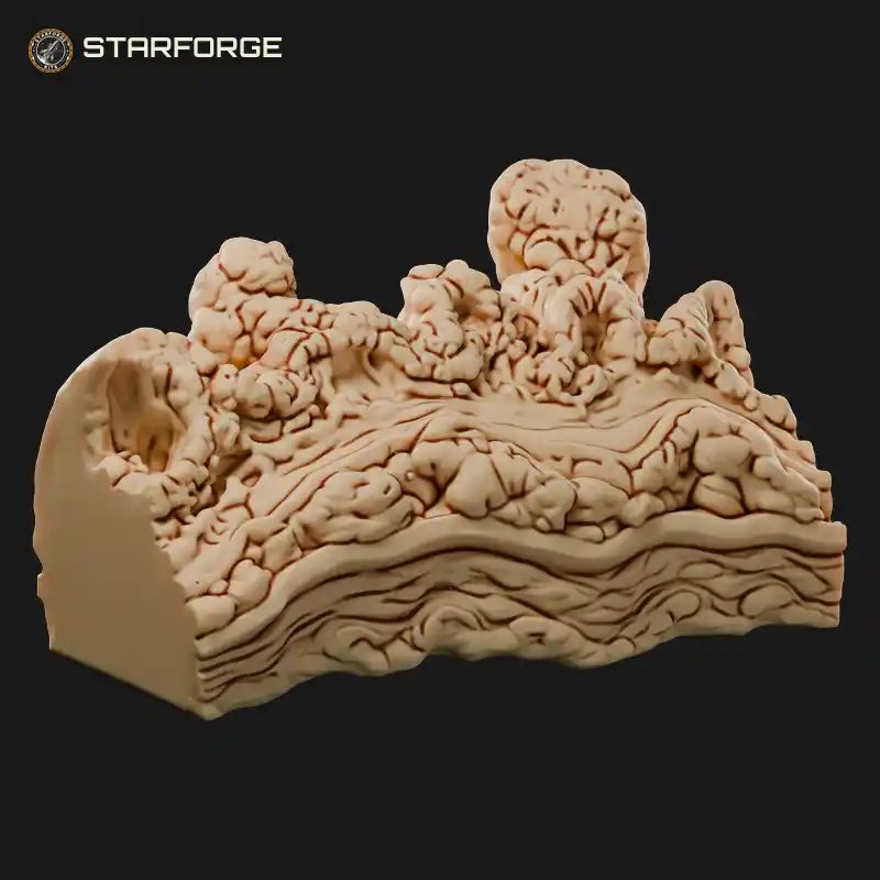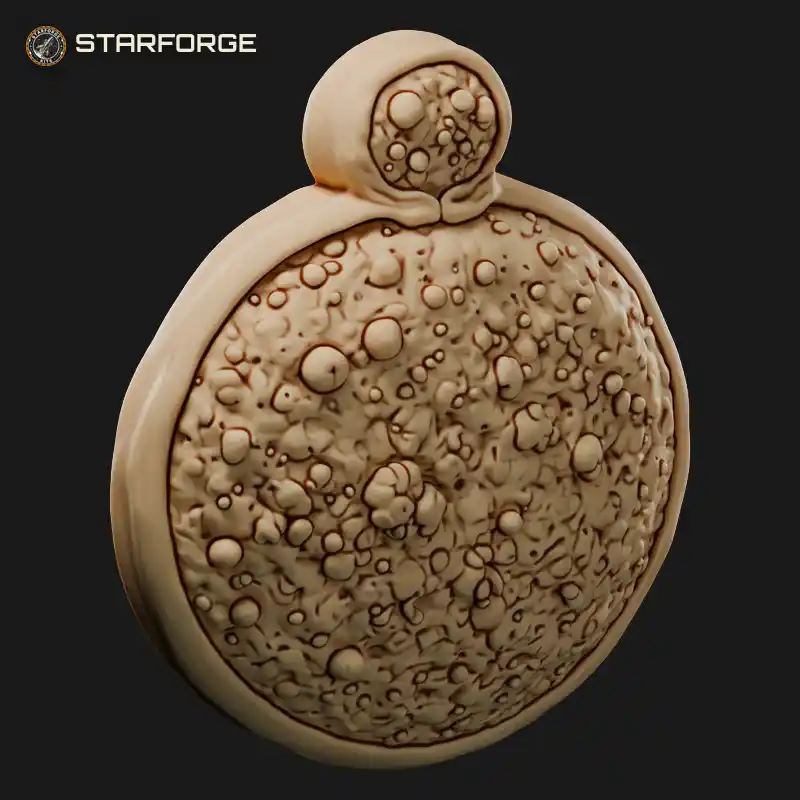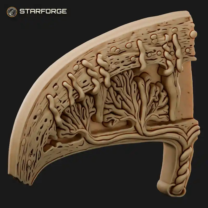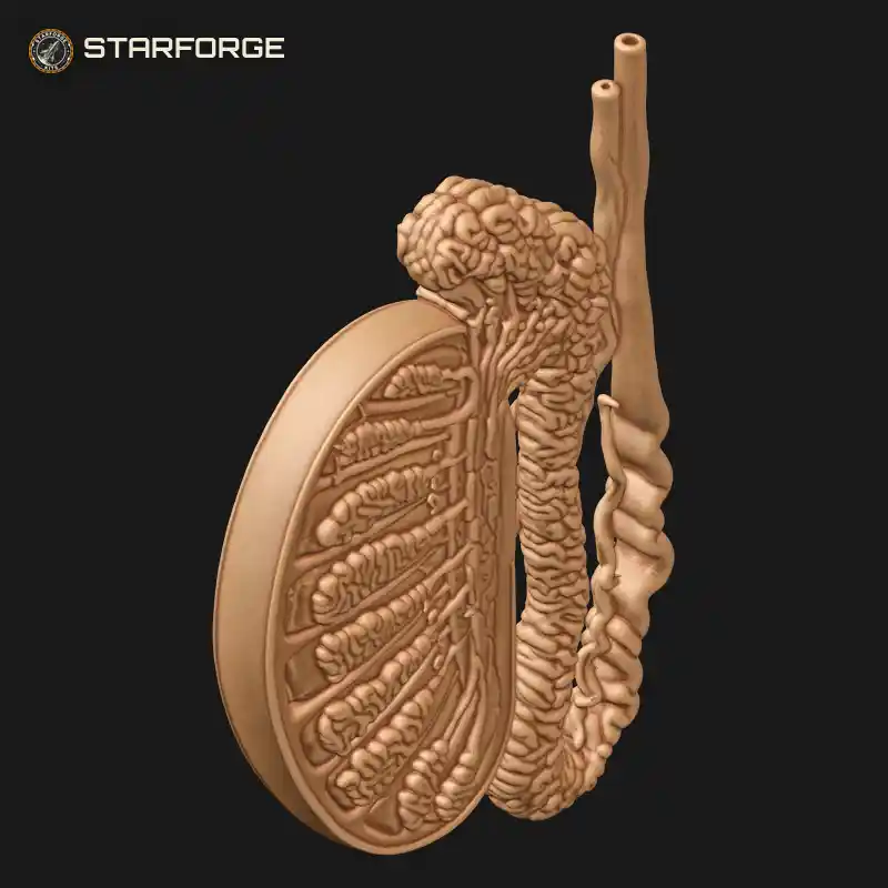
Testis Sagittal Cross Section with Septa Lobules Coiled Tubules Rete Testis Epididymis and Vas Deferens Education Model Realistic 3D Print STL File
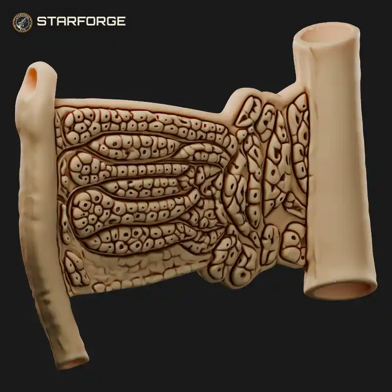
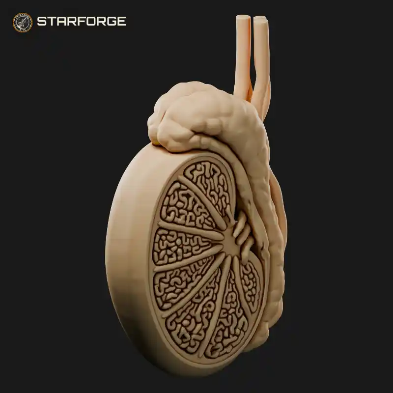
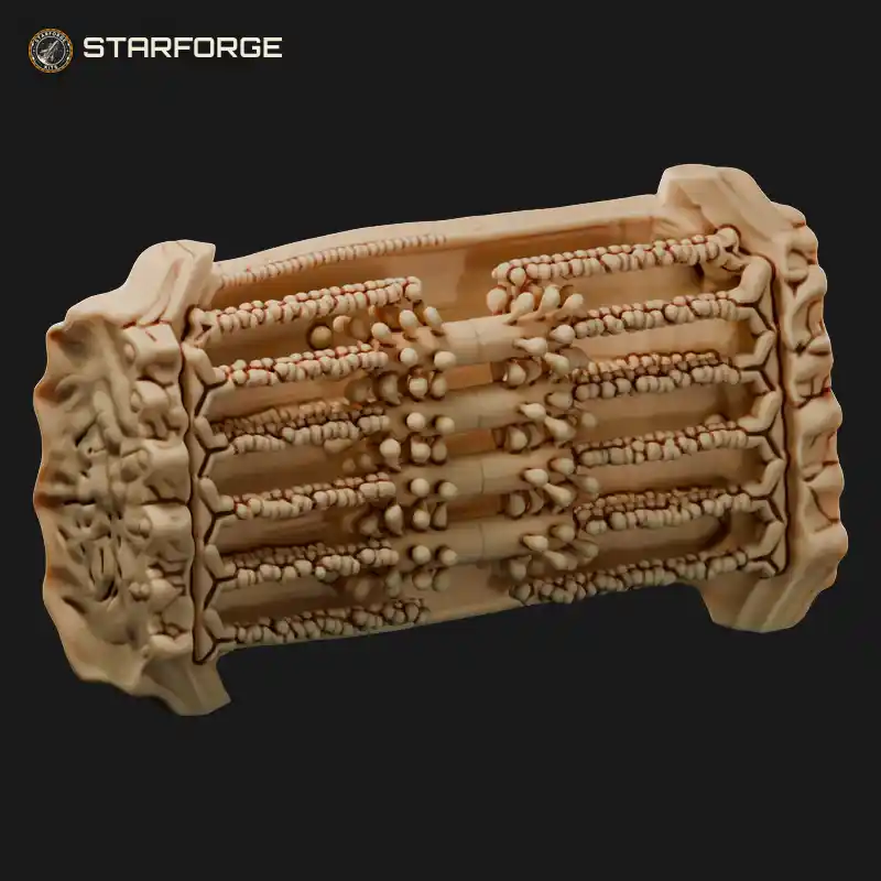
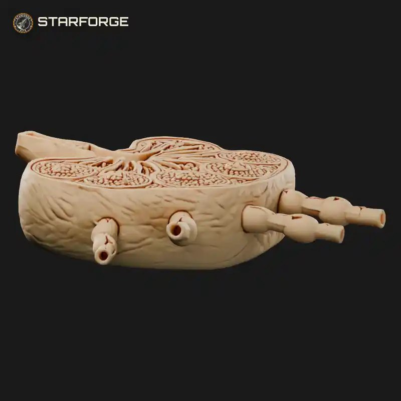
Sliced down the middle, this testis cross section opens like a disc to reveal the internal anatomy in clear relief. Thin septa divide the interior into wedge-shaped lobules, each packed with tightly coiled seminiferous tubules that drain toward the rete testis along the back edge. Alongside, the lumpy epididymis curls down the side and the vas deferens climbs upward as a slim, winding tube beside branching vessels. A solid teaching model for an anatomy class, a health sciences desk or a medical student studying male reproductive anatomy.
Related STL Files: