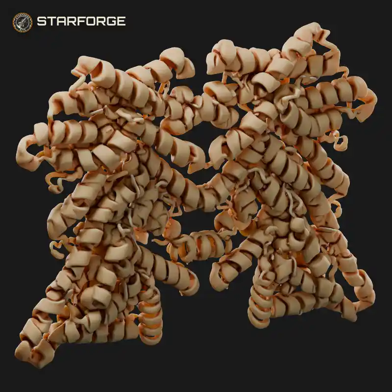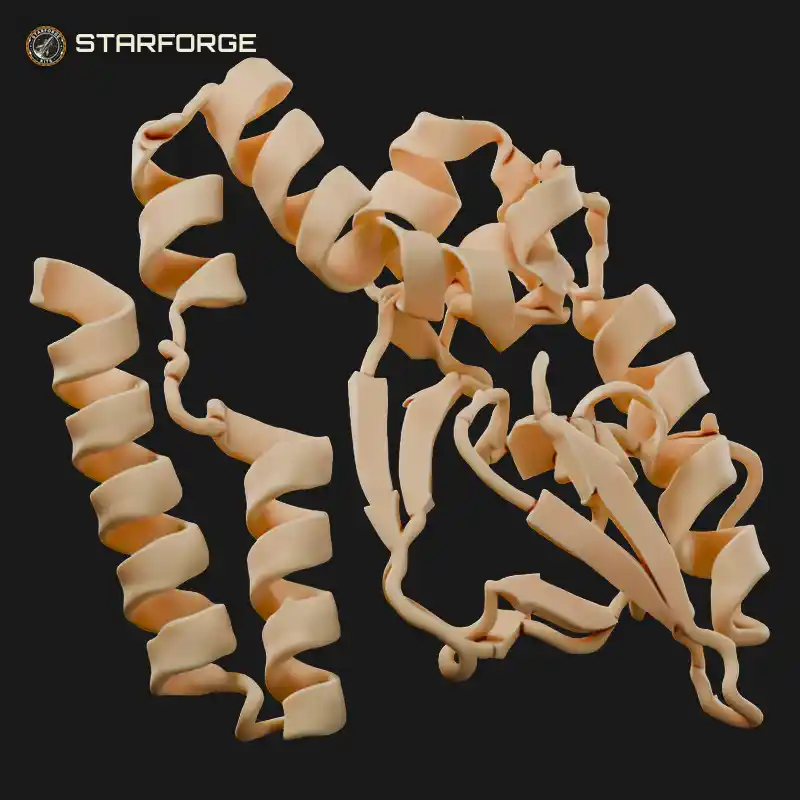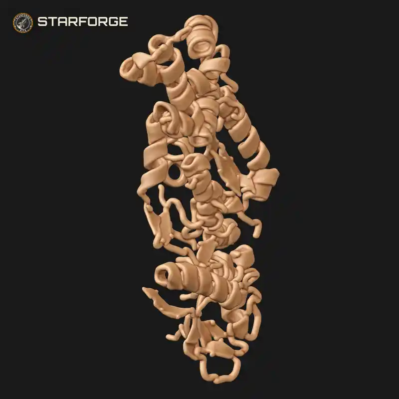
Whooping Cough Pertussis Toxin Protein with Enzymatic A Subunit Stacked on Binding B Oligomer Education Model Realistic 3D Print STL File
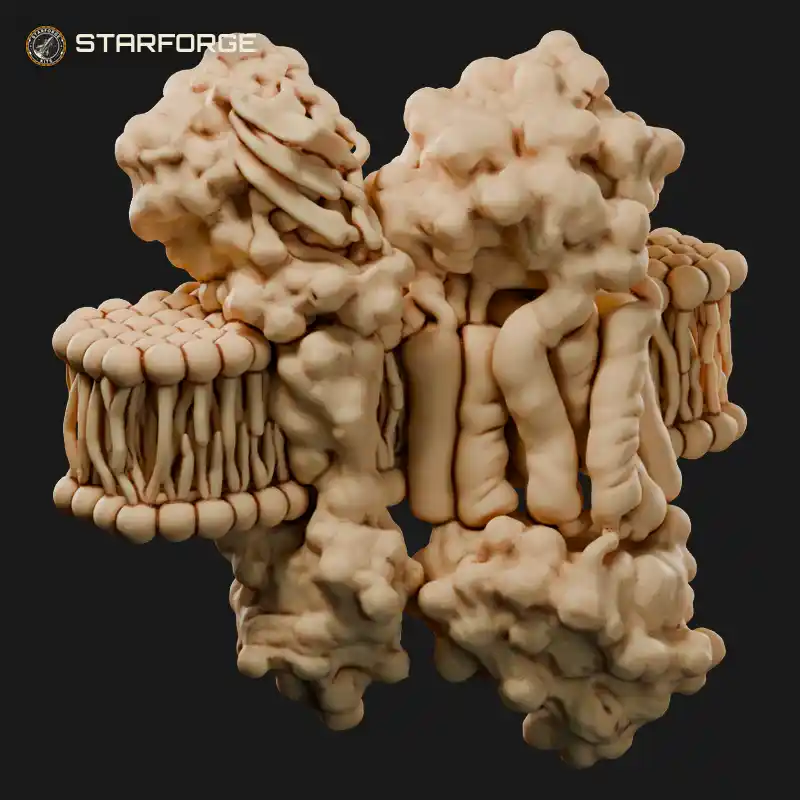
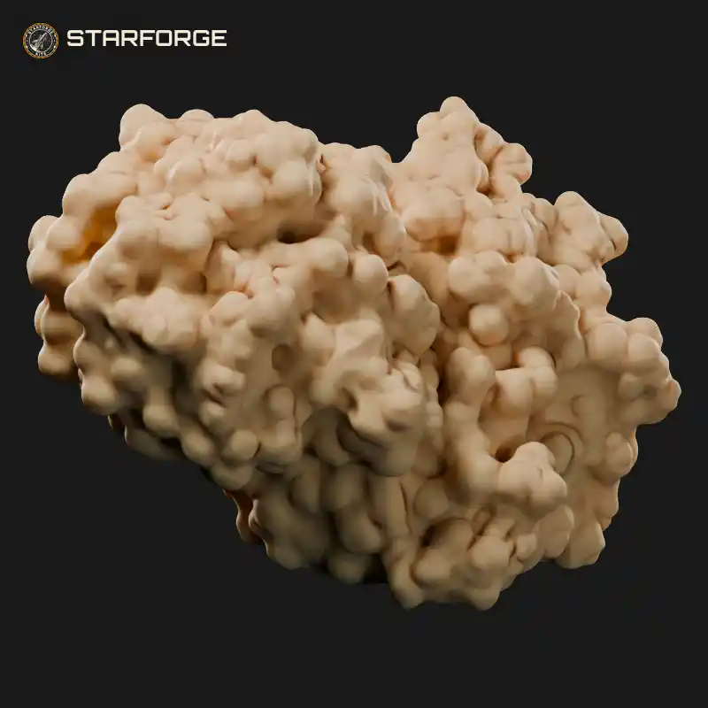
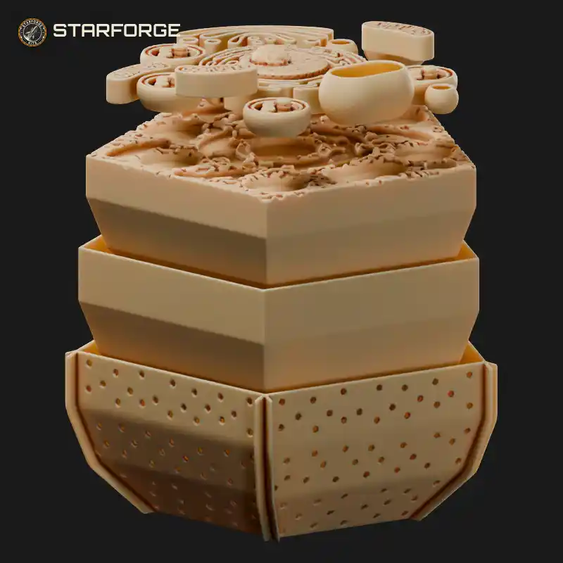
Ribbon-style helices and folded sheets twist together in this pertussis toxin protein, the molecule behind whooping cough, shown as a tall stacked structure. The upper enzymatic A subunit is a dense cluster of coiled spirals and loops, sitting on the ring-like binding B oligomer below with its own bundle of helices and arrow-shaped strands. Every ribbon stands open so the fold can be traced by eye. It suits a biochemistry classroom, a microbiology lab shelf or a display of protein structures for anyone curious about how bacterial toxins are built.
Related STL Files: