
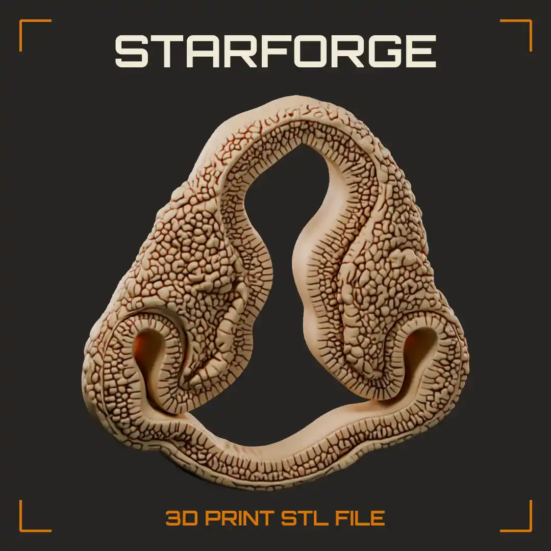
Embryo Head Cross Section with Midbrain Vesicle Paired Optic Vesicles and Thickening Lens Placodes Education Model Realistic StarForge 3D Print STL File
- Education Model
- Realistic
- Family Friendly
- Single Subject
- Display
- No Base
- Flat Panel
- Rough
- Flesh
- Biology
- Embryo
Related STL Files:

Cut straight through an early embryo head, this cross section shows the midbrain vesicle, paired optic vesicles and thickening lens placodes. The hollow neural tube rises in a tall arch, then curls out at each lower corner into a cupped optic vesicle tucked against the surface layer. Every wall is covered in fine cellular texture, so the layered tissue reads clearly from any angle. A strong teaching piece for embryology and developmental biology lessons on how the eye forms, and an unusual specimen for a science display shelf or lab bench.
Free StarForge 3D Viewer
Choose Your Membership
| Included each month | Starter | Merchant | Merchant+ | AI | AI Max |
|---|---|---|---|---|---|
| Credits | 45 | 150 | 450 | 1,500 | 4,000 |
| STL Downloads | up to45 | up to150 | up to450 | up to1,500 | up to4,000 |
| 3D Tools | Unlimited | Unlimited | Unlimited | Unlimited | Unlimited |
| AI 3D Models | up to4 | up to15 | up to45 | up to150 | up to400 |
| Sell 3D Prints | Included | Included | Included | Included | Included |
| Sign up |
Already a Member?LOGIN